
Hemocytometer (Counting of Cells)
Theory
For microbiology, cell culture, and many applications that require the use of cell suspensions, it is necessary to determine the concentration of cells. The device used for determining the number of cells per unit volume of a suspension is called a counting chamber. It is the most widely used type of chamber, originally designed for performing blood cell counts. Today, it is also used to count other types of cells and microscopic particles.
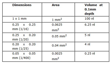
The hemocytometer was invented by Louis-Charles Malassez. It is a special type of microscope slide consisting of two chambers, each divided into nine large squares (1.0 mm x 1.0 mm), separated by triple lines. The area of each square is 1 mm². A cover glass is supported over the chambers at a fixed height of 0.1 mm, creating a known volume above the grid. Because of this, the entire counting grid lies within a volume of 0.9 mm³ on each side. The cell suspension is introduced beneath the cover glass. The hemocytometer is then placed on the microscope stage, and the cells are counted manually under magnification.
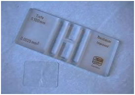
The glass slide has a rectangular indentation that creates an H-shaped chamber at the center. This chamber is engraved with a laser-etched grid of perpendicular lines. Two counting areas with ruled grids are separated by the horizontal bar of the "H". A very flat, reusable cover slip is used with the device. The cover slip is held precisely 0.1 mm above the counting surface by ground glass ridges on either side of the vertical grooves of the "H". Because the depth and the area of the squares are precisely known, the volume above each square is also known, allowing for accurate calculations of cell concentration.
To use the hemocytometer, the cover slip is placed on the device, and the chamber is filled with the cell suspension. There is a notch or V-shaped groove at each end of the chamber where the sample is introduced. The liquid is drawn into the chamber by capillary action. The cover glass does not float on the liquid but is held at a precise height. The grid’s arrangement of squares of different sizes allows for easy and systematic counting of cells. Using this method, it is possible to accurately determine the number of cells in a specified volume.
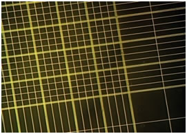
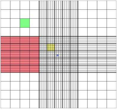
The ruled area of the hemocytometer consists of several large 1 × 1 mm (1 mm²) squares, which are subdivided in three ways: 0.25 × 0.25 mm (0.0625 mm²), 0.25 × 0.20 mm (0.05 mm²), and 0.20 × 0.20 mm (0.04 mm²). The central 1 × 1 mm square, which contains 0.20 × 0.20 mm subdivisions, is further divided into 0.05 × 0.05 mm (0.0025 mm²) squares.
Place the coverslip (0.1 mm thick) on the raised edges of the hemocytometer. This setup defines a specific volume above each square, allowing for accurate cell counting.